0 Medical One Liners 451-460


451. Periampullary carcinoma: Double duct sign

452. Carcinoma head of pancreas: Barium study - Widening of C loop of duodenum.

453. Down's syndrome: Antenatal ultrasound 
  • Increased Nuchal Translucency >3mm (between 11.3 to 13.6 weeks gestation)
    • Not to be confused with Nuchal fold thickness which is taken between 18 to 22 weeks of gestation
  • Absent / Hypoplastic Nasal bone
454. Ostium secundum is the Most common type of ASD, 
  • but in Down's syndrome - Ostium Primum.
455. Pancreatits: Investigation
  • Acute pancreatits: CECT is best
  • Chronic Pancreatitis: ERCP best, shows chain of lakes

456. Vertebral collapse
  • Normal IV disc
    • Trauma
    • Neoplasm
      • Adult - Multiple myeloma , Metastasis
      • Child - Histiocytosis
  • Reduced IV disc - Pott's spine
457. Most commonly PET scan uses 18 FDG
  • In myocardial perfusion - PET uses Rubidium 82
458. Bone infarct / Avascular necrosis - Best test is MRI

459. Vaginal epithelium is derived from endoderm of the urogenital sinus.

460. Half life of 18-FDG is 110 mins.


0 Renal Tuberculosis


Author: Dr. Ayush Goel


Renal tuberculosis occurs due to hematogenous spread.

Salient points:

  • Earliest symptom: Frequency of micturation
  • Lab reports: Sterile Pyuria
  • Best Radiological test: IVP
    • Earliest feature - feathery appearance of calyces
    • Late - Putty Kidney/Cement Kidney (Autonephrectomy)
      • Note: Autosplenectomy is seen in Sickel cell disease
    • Urinary bladder - Thimble bladder (thick wall and low capacity)
    • Ureter - Golf hole ureters
Note: Bladder calcification is uncommon in TB. Bladder calcification is seen in Schistosomiasis.


0 Radiology Quiz 6


Author: Dr. Ayush Goel


Q. What is the diagnosis of this classical appearance of lower limb during antenatal ultrasound scan ?

Image courtesy: Dr Ayush Goel


ANSWER:
Answer: Congenital Talipes Equino Varus (CTEV / Club Foot)


0 Radiology Quiz 5


Author: Dr. Ayush Goel


Q. Whats the diagnosis for this classical appearance ?

Image courtesy: Dr Ayush Goel

ANSWER:
Answer: Rickets - Cupping and flaring of metaphysis with widening of growth plate.


0 Medical One Liners 441-450


441. MRI - Gadolinium contrast is used (when needed). In patients with renal failure, it may cause Nephrogenic Systemic Fibrosis - plaques of skin (firm, indurated and erythematous) with subcutaneous oedema.

442. Air bronchograms can be seen in alveolar pathologies:
  • Pneumonia
  • Pulmonary edema
  • Hyaline membrane disease
  • Bronchoalveolar carcinoma
443. Pneumatocele can be seen in
  • Staphylococcal and klebsiella pneumonia
  • Hydrocarbon poisoning
444. Viral and mycoplasma pneumonia gives interstitial pattern. No alveolar exudates are seen.

445. Hydatid cyst:
  • Ultrasound: Cart wheel appearance
  • in Lungs it does not calcify
  • Chest X-ray: if contained rupture - water lily sign
    • Note: Drooping lily sign is seen on IVP in duplex collecting system with upper moiety obstruction
446. Egg in cup:
  • calyces: Papillary necrosis
  • heart: constrictive pericarditis
447. Focal nodular hyperplasia (FNH) has high Kupffer cells and takes up technetium 99m sulphur colloid.

448. In Blunt trauma cases:
  • Ideal and gold standard investigation: Contrast enhanced CT (CECT)
  • If haemodynamically unstable/1st Investigation: FAST (Focused assessment sonography in trauma)
449. Beaded appearance in Renal artery/Internal carotid/External Iliac: Fibromuscular dysplasia

450. Only Veins (hepatic and portal) form basis of division of couinaud segments of the liver.


2 Pulmonary sequestration


Author: Dr. Ayush Goel


Pulmonary Sequestration = accessory lung (embryonic cut off of a segment from main lung)

 Types:
Intralobar
Extralobar

More common (75-85% cases)
Less common (15-25% cases)
 Presentation
Usually Child with recurrent infections
Usually Neonate with respiratory distress and cyanosis
Arterial supply
Aorta (Same in both types)
Venous supply
usually Pulmonary venous  drainage
Usually systemic venous drainage
Pleura
Same pleura as ipsilateral lung
Separate pleura from ipsilateral lung
Site
60% cases affect left lower lobe (Most commonly Posterior basal segment); 40% right lower lobe.
Always affect left lower lobe; can be subdiaphragmatic in 10% cases

Sequestrated lung appears hyperechoic than rest of lung on prenatal ultrasonography


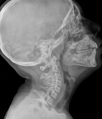
1 Radiology Quiz 4


Author: Dr. Ayush Goel


Q. Whats your diagnosis ?

Image courtesy: Dr Ayush Goel and Dr Subhash Goel


ANSWER:
Answer: Enlarged Adenoids and Tonsils


Related Posts Plugin for WordPress, Blogger...
To Search for a "particular word" on "the page on your screen" , press Ctrl + F and then type the word you need to search on the visible page.

eg: If you need to search the word "Anatomy" on this page -- Press "Ctrl + F" , (a box will appear) and then type Anatomy in the box that has appeared.